Cardiovascular Imaging
Ohan Cardiovascular Innovations provides researchers with a non-invasive anatomical image of heart, blood vessels, tumors or normal abdominal organs with up to 50uM resolution. The available ultrasound system’s features linear array ultrasound transducer capable of imaging in the 25-75MHz range, allowing multiple focal depths and improved feature detection.
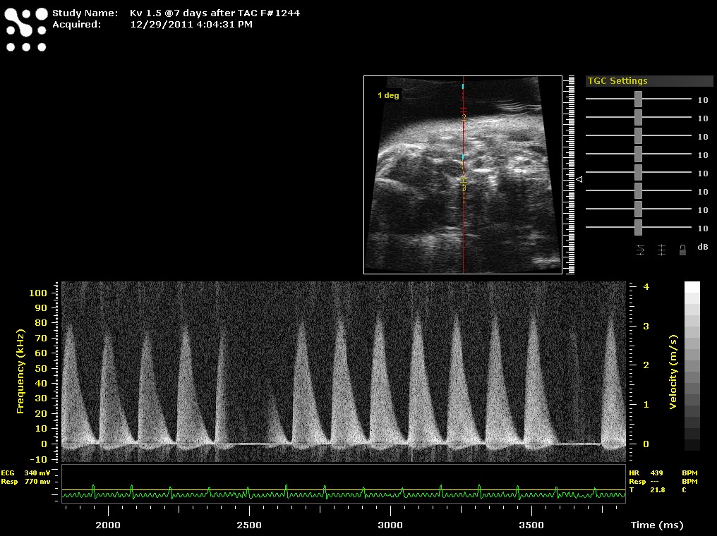
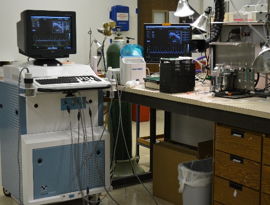
Transthoracic Echocardiography – we will be using the Vevo 2100 and Vevo 770 high frequency ultrasound systems for cardiovascular imaging. All images will be done under isoflurane anesthesia. If necessary, we will modify the type of anesthesia if isoflurane can have an affect on your research results.
Non-invasive Transthoracic Cardiac Ultrasound
- 2D and basic mode – Parasternal short and long axis 2D video images will be recorded and saved. This mode permits the measurement of endocardial and epicardial surface area, surface area changes in normal the heart and after cardiac remodeling. Ejection Fraction (%EF), Fractional Area Changes (%FAC), and left ventricular mass (LAV mass by mg) can be calculated by using this mode.
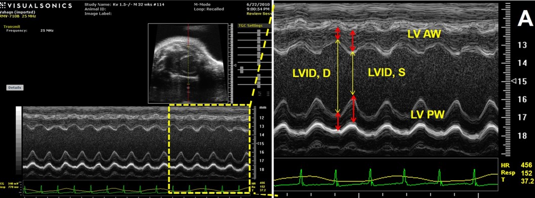
- M-mode – recordings, M-mode at mid-papillary muscle level from short and long axis pictures. 3-5 different images will be recorded.
Utilizing M-mode images we will measure:
- Left ventricular (LV) volumes (LVvold and LVvols)
- Stroke Volume (SV)
- Cardiac Output (CO)
- Calculated % Ejection Fraction (%EF)
- LV Internal Diameter (LVIDd and LVIDs)
- Calculated % Fractional Shortening (% FS)
- Wall thicknesses,
- LV mass

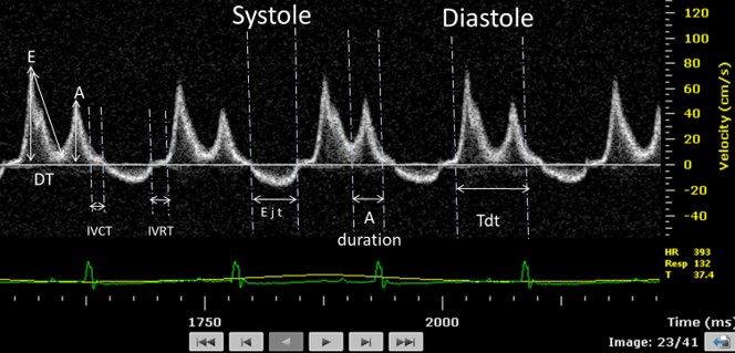
- Doppler Imaging - This method is used to detect moving blood cells or other moving structures (myocardial tissue) and measure their direction and speed of movement.
- E peck velocity
- A peak velocity
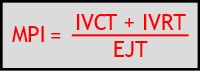
- Isovolumetric contraction time ( IVCT)
- Isovolumetric relaxation time (IVRT)
- Ejection time(ET)
- Myocardial performance index (MPI)